Figure 5.
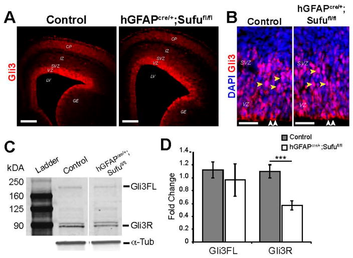
Reduced Gli3R levels in the VZ/SVZ of the E16.5 hGFAPcre/+;Sufufl/fl neocortex. (A,B) Gli3 immunostaining showed highly specific expression in the VZ/SVZ of the E16.5 neocortex of control and hGFAPcre/+;Sufufl/fl mice (A); Confocal z-stack imaging showed that the subcellular localization of Gli3 did not significantly differ between control and hGFAPcre/+;Sufufl/fl VZ/SVZ, where Gli3 appeared to be cytoplasmic along the ventricular lining (white arrows) and nuclear localized in the SVZ (yellow arrows) (B); Scale bars = 200 μm in (A); 50 μm in (B); GE, ganglionic eminence; (C) Western blot analysis of protein extracts from the E16.5 neocortex showed the presence of both Gli3FL and cleaved Gli3R proteins, with Gli3R being predominant in both control and hGFAPcre/+;Sufufl/fl mice. However, quantification of Gli3FL and Gli3R (D) showed significantly reduced Gli3R protein levels in the hGFAPcre/+;Sufufl/fl neocortex compared to controls. α-Tubulin (α-Tub) was used as a loading control; *** p-value < 0.01; Gli3FL, full-length Gli3; Gli3R, cleaved Gli3R repressor.

