Figure 8.
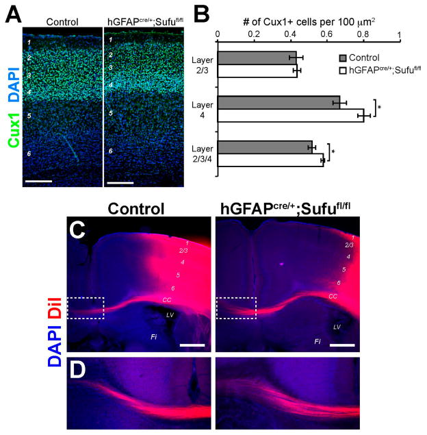
Mild increase in Cux1+ projection neurons and normal callosal projections in the postnatal hGFAPcre/+;Sufufl/fl neocortex. (A,B) Cux1 immunolabeling of upper layer projection neurons showed a comparable distribution of Cux1+ neurons, with a densely packed layer 4, in the P7 hGFAPcre/+;Sufufl/fl neocortex (A); Quantification of Cux1+ projection neurons (B) yielded comparable densities of Cux1+ neurons in Layer 2/3 between control and hGFAPcre/+;Sufufl/fl neocortex. A borderline increase in the density of Cux1+ neurons were quantified in layers 2–4 of the P7 hGFAPcre/+;Sufufl/fl neocortex (p-value = 0.054) and appears to be due to a mild increase in the density of Cux1+ neurons in layer 4 (p-value = 0.058). Scale bars = 200 μm. CC, corpus callosum; LV, lateral ventricles; FI, fimbria; (C,D) DiI tracing showed that callosal projections that originate from upper cortical layers were grossly unaffected in the P15 hGFAPcre/+;Sufufl/fl neocortex and is comparable with control littermates (C); Callosal projections were able to project across the midline into the contralateral hemisphere (boxed, D); Scale bars = 500 μm. DiI, 1,1′-dioctadecyl-3,3,3′,3′-tetramethylindocarbocyanine.

