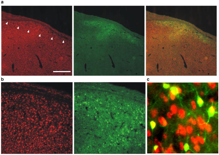
Fig. 4.

AAV hSyn1 GFP transduces HVC neurons. Photomicrographs show NeuN-positive neurons (red; right panels), GFP reporter signals (green; middle panels) and merged images (left panels). a Low magnification images show HVC (arrowheads denote ventral border) as the slight increase in neuronal density lying just superficial to the thin hyperpallial layer. Scale bar 500 μm. b Higher magnification is shown in left and middle panels. c Merge of even higher power image illustrates that only NeuN positive cells are transduced
