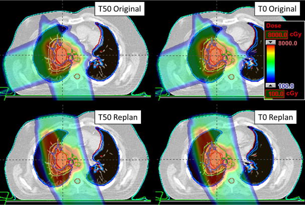
FIG. 6.

An example of the ΔWET guided plan (bottom) compared to the original, clinical plan (top). An axial slice plan dose is shown on the top for T50 (left) and T0 (right) image set. The ΔWET guided plan has new field angles of 155° and 350° which were determined using the ΔWET analysis program.
