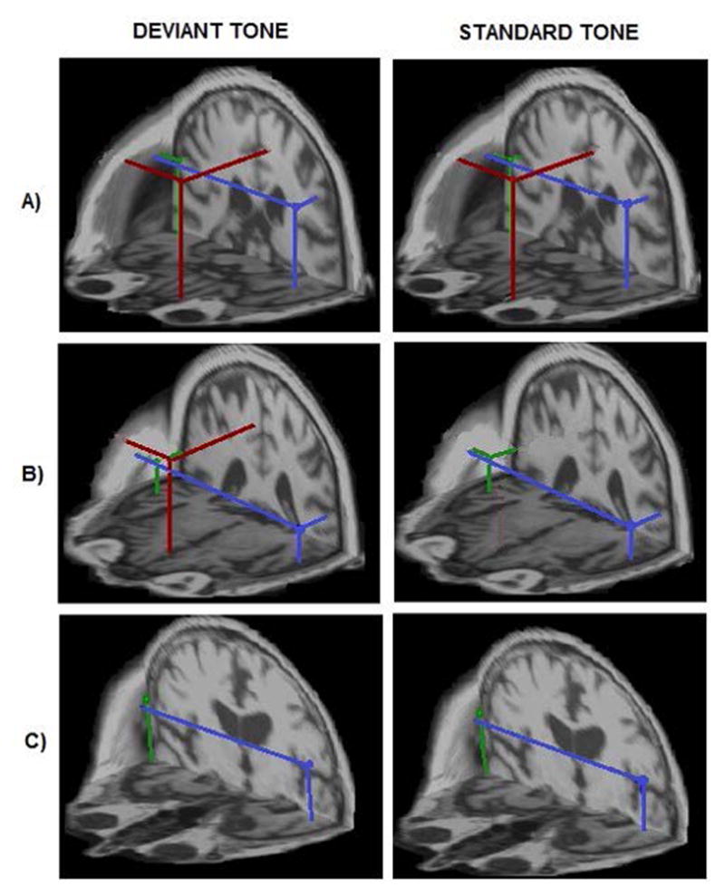
Fig. 1. Three distinct types of auditory gating network identified in healthy controls (a), possible preclinical AD (b) and symptomatic AD patients (c).

Localization of auditory gating generators estimated in the 30–100 ms time interval evoked by the tones of an oddball paradigm in three representative subjects across conditions. The best-fitting source locations are superimposed on individual volumetric MRI head data to achieve a spatial (3D) rendering of the auditory gating topology. Panel A) shows the healthy gating topology type where all three gating generators, mPFC (red dot) in addition to bilateral STG sources (green and blue dots; right and left STG generators, respectively), were active in processing both tone conditions (4/10 controls). Panel B) shows an altered gating topology type characterized by selective mPFC activation only by the deviant tone (6/10 controls and one aMCI). Panel C) shows the third topology type where the mPFC generator for both standard and deviant tones was missing. This type of network was found in symptomatic AD patients.
