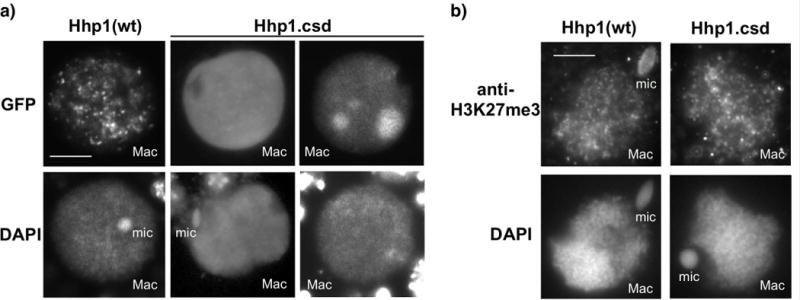
Fig. 5.

Loss of the CSD reduces chromatin body targeting of Hhp1 in nuclei from growing cells. a) Cells were induced to express GFP-Hhp1Δcsd (“Hhp1.csd”), then stained with DAPI and visualized by epifluorescence microscopy. Two sets of nuclear images representing two observed localization patterns of GFP-Hhp1Δcsd are shown. Scale bar = 5μm shown in first panel applies to all images. b) Cells expressing GFP-Hhp1Δcsd were subjected to immunofluorescence with anti-H3K27me3, counterstained with DAPI, and visualized by epifluorescence microscopy. “Mac”, macronucleus; “mic”, micronucleus. Scale bar = 5μm shown in first panel applies to all images.
