
Figure 1. Downregulation of NELF protein during granulocytic differentiation.
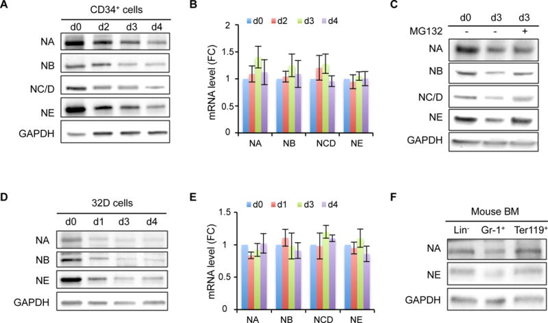
(A) Western blot of NELF subunits from different days during granulocytic differentiation of human CD34+ HSPCs. GAPDH serves as a loading control. NA: NELF-A, NB: NELF-B, NC/D: NELF-C/D, NE: NELF-E. Quantification results are in Figure S1C.
(B) Quantitative RT-PCR of mRNA levels of NELF genes during granulocytic differentiation of CD34+ cells. Gene expression is normalized to β-actin and presented as fold change relative to d0 (n=3, mean ± SEM).
(C) Western blot of NELF subunits on differentiation day 3. MG132 treatment was done by adding 0.2uM MG132 for overnight before harvesting cells for protein extraction. Quantification results are in Figure S1D.
(D) Western blot of NELF subunits during neutrophil differentiation of mouse 32Dcl3 cell line. Quantification results are in Figure S1E.
(E) Quantitative RT-PCR of mRNA levels of Nelf genes during neutrophil differentiation of mouse 32Dcl3 cells. Gene expression is normalized to β-actin and presented as fold change relative to d0 (n=3, mean ± SEM).
(F) Western blot of NELF-A and -E subunits from cells isolated from mouse bone marrow.