
Figure 1.
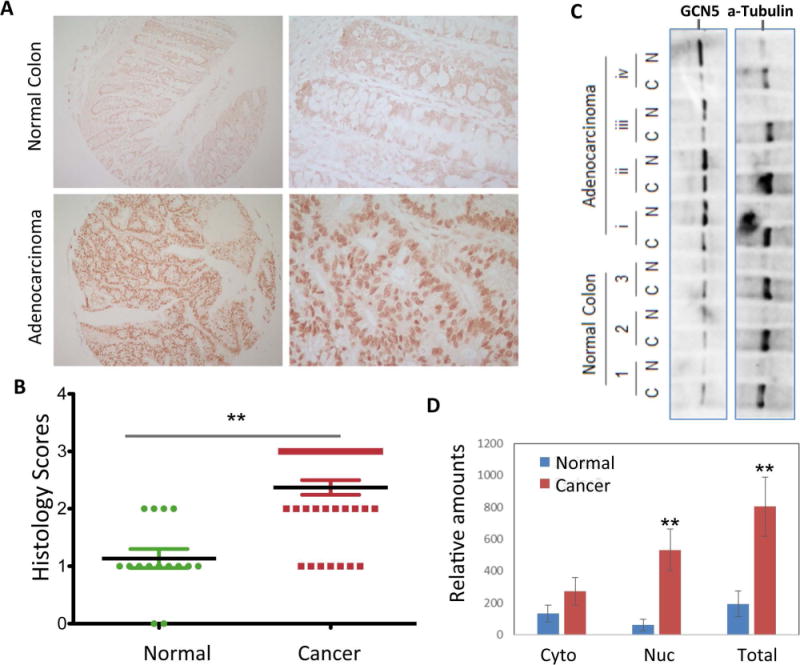
Elevated GCN5 expression in human colon cancer tissues. (A, B) The human colon carcinoma tissues were used for the immunohistological staining with GCN5-specific Abs. Normal adjacent tissues were used as controls. (A) Representative images are shown and (B) the histological scores from 120 adenocarcinoma and their adjacent control tissues are indicated. Student’s t-test was used for the statistic analysis. (C, D). Subcellular fractionation of freshly collected human colon cancer tissues and normal controls was performed, the expression levels of GCN5 and the cytoplasmic protein α-tubulin as a control.