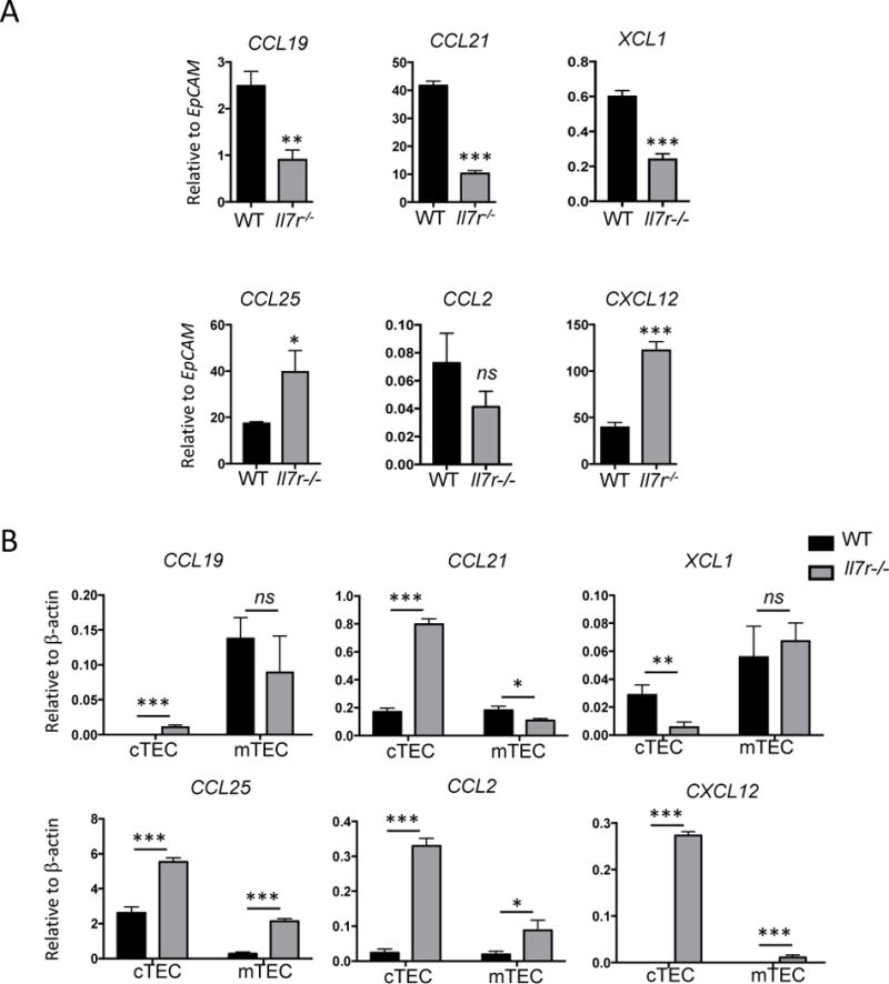
Figure 4.

Perturbation in chemokine expression in the Il7r−/− thymus. A. Whole homogenized thymus was used as a template for qRT-PCR to measure the amount of chemokine mRNA produced in the adult WT versus Il7r−/− thymus. To account for the 5-fold greater number of TECs in the Il7r−/− thymus versus the WT thymus, results were normalized to EpCAM mRNA levels. B. TECs were sorted based on expression of EpCAM, and Ly51 (cTECs) or UEA-1 (mTECs). cDNA was generated from sorted TEC subsets and used as template for qRT-PCR. n=3. Values were normalized to β-actin mRNA levels. Data is representative of at least two separate experiments. Graphs depict means +/− SEM. Statistical significance was calculated using a t-test; *** p<0.005, **p<0.01, *p>0.05.
