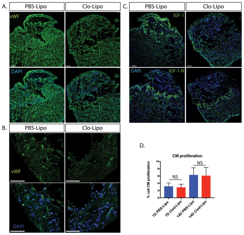
Figure 5. Blood vessel density and IGF signaling in macrophage depleted animals is altered in Clo-Lipo animals where cardiomyocyte proliferation appears unchanged.
A. Endothelial cell staining using von Wilebrand Factor (vWF) demonstrated a reduced vascular network in Clo-Lipo animals at 14dpi. B. higher magnification of vWF staining at wound margin showed reduction in blood vessel outgrowth. C. Insulin-like Growth Factor (IGF-1) ligand expression was downregulated in Clo-Lipo injured hearts at 14dpi with weak epicardial expression maintained. Conversely, IGF-1-Receptor expression was upregulated specifically in cardiomyocytes (CMs) at the wound margin and maintained in Clo-Lipo treated animals. D. CM proliferation was measured using PCNA+ Nkx2.5+ cells on 7dpi and 15 dpi hearts and no significant difference was detected. CM proliferation measured using at least 3 sections per animal and 3 animals per time-point. Scale bar = 100μM.

