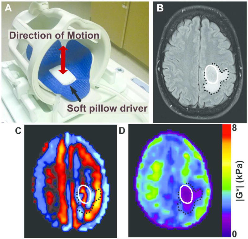
Figure 1.
Brain MRE experimental setup and image procesing. (A) Brain MRE soft pillow driver placed within the 8-channel MRI head coil and positioned beneath the head to induce shear waves into the brain. (B) Axial T2 FLAIR image of a glioblastoma, IDH1 wild-type (male, age 51) with tumor denoted by solid white line and peritumoral edema by black dotted line. (C) MRE shear wave image and (D) elastogram or stiffness map displaying a soft tumor with a stiffness of 1.1kPa in the tumor compared to 3.5 kPa in a size-matched region of unaffected white matter on the contralateral hemisphere.

