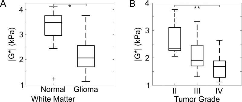
Figure 2.
(A) Gliomas were softer than normal brain tissue, compared to size-matched regions of interest in the unaffected contralateral white matter (*p < .001). An outlier is indicated by a plus sign, and whiskers on the boxplot indicate the 25th and 75th percentiles. (B) Glioma stiffness decreased with increasing tumor grade (**p < .05).

