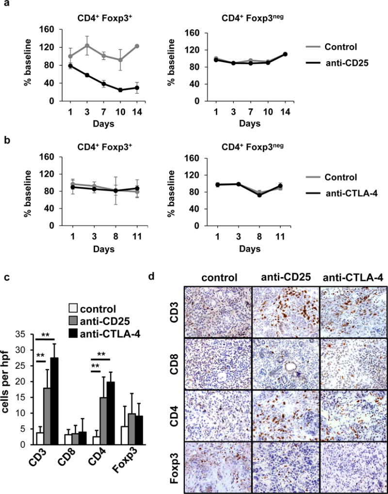
Fig. 2. Antibodies targeting CD25 and CTLA-4 induce CD4+ T cell infiltration into spontaneous PDAC tumors.

Mice (n=4/group) were treated with a, anti-CD25 (day 1) versus isotype control (day 1) or b, anti-CTLA-4 (days 1, 4, 8, 11), or isotype control (days 1, 4, 8, 11). Shown is the impact of treatment with a, anti-CD25 (black) versus control (grey) and b, anti-CTLA-4 (black) versus control (grey) on the frequency of CD3+CD4+Foxp3+ Tregs and CD3+CD4+Foxp3neg conventional T cells detected in the peripheral blood after treatment. Shown is c, quantification per high power field (hpf) and d, representative images of immunohistochemistry to detect CD3, CD8, CD4 and Foxp3 cells in PDAC tumors from KPC mice at 14+2 days after beginning treatment with anti-CD25 (n=5) or anti-CTLA-4 (n=5) with comparison to isotype control (n=4). Scale at 40× magnification.
