
Figure 8.
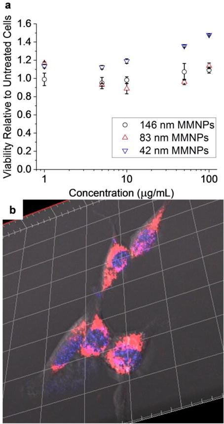
In vitro investigation of MMNP-cell interactions. (a) MMNP cytocompatibility with NIH/3T3 fibroblasts as measured by neutral red uptake viability assay. Error bars represent standard errors of triplicate experiments. (b) Representative confocal microscopy three-dimensional (3D) z-stack reconstruction image of Hoechst-stained NIH/3T3 fibroblasts treated with 20 μg mL−1 MMNP@PEG@RA123. Hoechst stain (blue) and rhodamine fluorescence (red/pink) are shown here; Scale bar: 20 μm between gridlines.