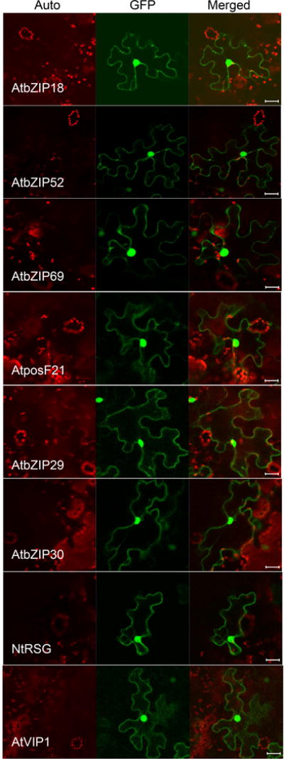
Fig. 5.

AtVIP1and its homologs localize to the cell nucleus and cytoplasm. The indicated proteins tagged with GFP were transiently expressed in agroinfiltrated leaf epidermis of N. benthamiana, and analyzed by confocal microscopy three days post-infiltration. GFP signal is in green, plastid autofluorescence is in red. Images are single confocal sections, representative of images obtained in two independent experiments performed for each protein; for each experiment, three infiltrations were performed on three different leaves, with two images recorded per infiltration. Scale bars= 20 μM.
