Fig. 7.

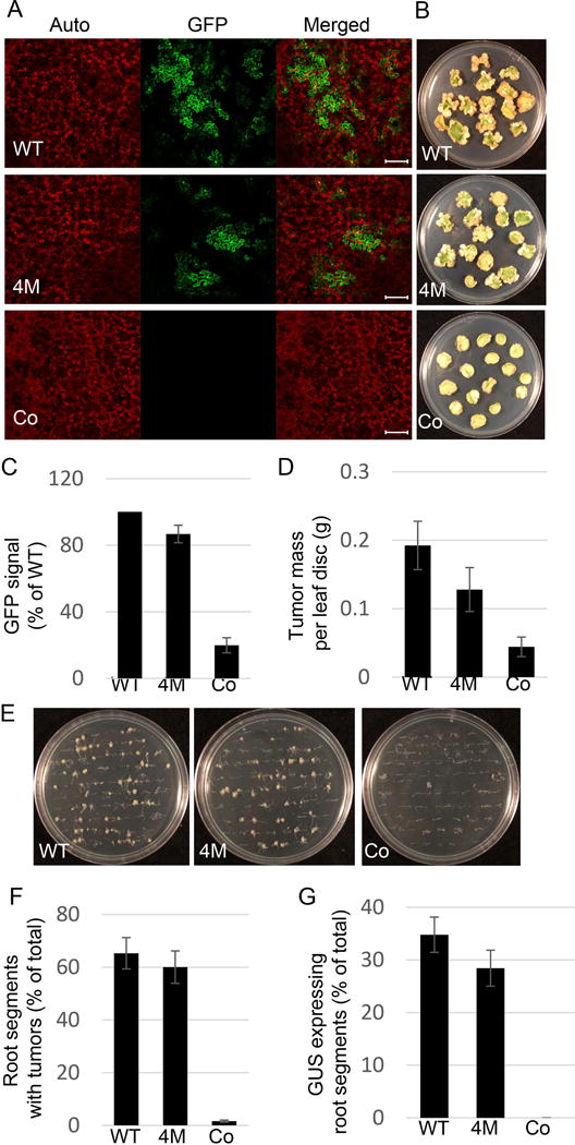
The effect of C58 VirE2 4M on transient and stable transformation capacity of Agrobacterium. (A) Transient transformation of N. benthamiana leaves. Plant tissues were coinfiltrated with two cultures of the Agrobacterium strain mx358 that lacks its endogenous virE2 gene: one harboring a plasmid that expresses either wild-type C58 VirE2 (pEpA6-C58virE2) or C58 VirE2 4M (pEpA6-C58virE2–4M) in the bacterium, or carries an empty plasmid (pEpA6), and the other harboring a binary plasmid that expresses the GFP reporter. Three days post-infiltration, GFP expression in the inoculated tissues was analyzed by confocal microscopy. GFP signal is in green, plastid autofluorescence is in red. Images are single confocal sections, representative of images obtained in two independent experiments performed for each protein; for each experiment, three infiltrations were performed on three different leaves, with two images recorded per infiltration. Scale bars= 100 μM. (B) Stable transformation of N. tabacum leaves. Leaf discs were inoculated with the Agrobacterium strain mx358 with a plasmid that expresses either wild-type C58 VirE2 (pEpA6-C58virE2) or C58 VirE2 4M (pEpA6-C58virE2-4M), or carries an empty plasmid (pEpA6), and the tumors were photographed three weeks after inoculation. Three plates, each containing 15 leaf discs, were used for each condition. (C) Quantification of GFP expression shown in (A). GFP signal was quantified as described in Fig. 4. Signal obtained with wild-type C58 VirE2 was set as 100%. Error bars represent SEM of N=3 independent biological replicates (leaves). (D) Quantification of tumor formation shown in (B). Tumors were scored by their mass. Error bars represent SEM of N=3 independent biological replicates (plates). (E) Stable transformation of A. thaliana roots. Root segments were inoculated with the Agrobacterium strain mx358 with a plasmid that expresses either wild-type C58 VirE2 (pEpA6-C58virE2) or C58 VirE2 4M (pEpA6-C58virE2-4M), or carries an empty plasmid (pEpA6), and the tumors were photographed three weeks after inoculation. Three plates, each containing 50 root segments, were used for each condition. (F) Quantification of tumor formation shown in (E). Roots segments with tumors were counted and expressed as percent of the total inoculated roots. Error bars represent SEM of N=3 independent biological replicates (plates). (G) Transient transformation of A. thaliana roots. Root segments were inoculated with two cultures of the Agrobacterium strain mx358: one with a plasmid expressing wild-type C58 VirE2 (pEpA6-C58virE2) or C58 VirE2 4M (pEpA6-C58virE2-4M), or carries an empty plasmid (pEpA6), and the other harboring a binary plasmid expressing the GUS reporter. Three days post-inoculation, GUS activity in the inoculated tissues was analyzed by histochemical staining, and the number of stained roots counted and expressed as percent of the total inoculated roots. Three plates, each containing 50 root segments, were used for each condition. WT, wild-type C58 VirE2; 4M, C58 VirE2 4M; Co, empty vector.
