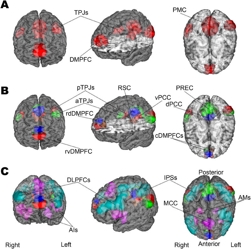
Figure 1. Target network definitions.
The regions of interest (ROIs) are rendered on the MNI standard brain with frontal, diagonal, and top views. A The DMN is represented by 4 ROIs, according to how the main network nodes are frequently studied in neuroimaging research. These comprise the dorsomedial prefrontal cortex (DMPFC), posteromedial cortex (PMC), and right/left temporoparietal junction (TPJ). B The DMN nodes are subdivided into 12 ROIs accounting for the distinct subnodes in the DMN that were recently established (Bzdok et al., 2016a; Bzdok et al., 2015; Bzdok et al., 2013; Eickhoff et al., 2016). According to this prior work, the functional core of the DMN (“DMN proper”) likely corresponds especially to its blue and red subnodes (the ventral and the dorsal PCCs, the left and right posterior TPJs, and the rostroventral and rostrodorsal DMPFC). C The DMN subnodes are supplemented by 9 ROIs for the dorsal attention network (DAN, light green) and saliency network (SN, purple), drawn from published quantitative meta-analyses (Bzdok et al., 2012; Rottschy et al., 2012). The DAN was composed of the dorsolateral prefrontal cortex (dlPFC) and intra-parietal sulcus (IPS) bilaterally. The SN included the midcingulate cortex (MCC) and the bilateral anterior insula (AI) as well as amygdala (AM). NeuroVault permanent link to all ROIs (21 in total) used in the present study: http://neurovault.org/collections/2216/.

