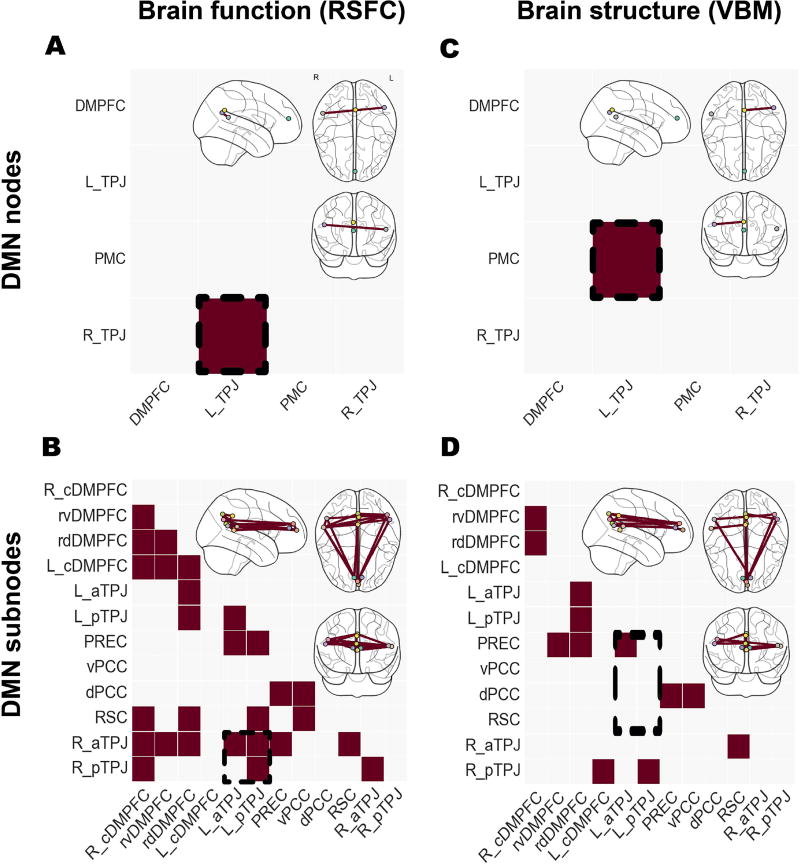
Figure 3. Studying node versus subnodes in the default mode network.
Significant differences in functional connectivity (left column, resting-state functional connectivity [RSFC]) and structural co-occurrence (right column, voxel-based morphometry [VBM]). Schizophrenic patients and healthy controls were compared based on the usual DMN nodes (upper row) and the topographically more fine-grained DMN subnode atlas (lower row). Richer brain signals have been captured by the recent parcellation of the DMN nodes, resulting in a higher number of statistically significant group effects. Analysis approaches based on collapsed DMN nodes may therefore obfuscate disease-specific patterns in fMRI signals as indexed by resting-state connectivity and in MRI signals as indexed by voxel-based morphometry. The glass brains were created using the nilearn Python package (Abraham et al., 2014).

