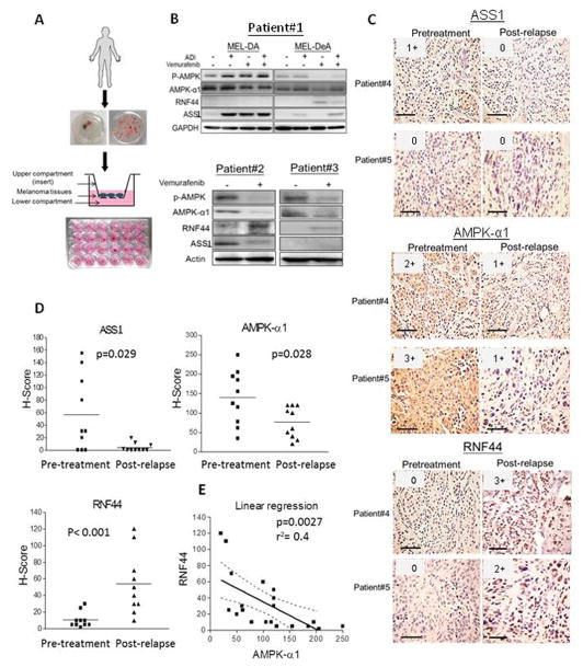
Fig. 7.
Attenuated ASS1 and AMPK-α1 but increased RNF44 levels were seen in tumors from BR/BMR patients. (A) The explant assay was shown as schematic workflow which has been described in section 2.14. (B) Tumor explants from melanoma patients (patient #2 and #3) who relapsed following treatment with vemurafenib were placed in the inserts and exposed to drugs (in lower compartments) for 48–72 hr. The cell lysates were subjected to immunoblot analysis. MEL-DA and MEL-DeA cells were primary cultures isolated from a melanoma patient #1, and represent pretreatment and BRAFi resistance (post-relapse), respectively. (C) ASS1, AMPK-α1, and RNF44 expressions in paraffin-embedded tumor tissues of patient #4 (resistant to BRAFi) and patient #5 (resistant to BRAFi/MEKi) were determined by IHC staining (scale bar = 100 μm). The intensities were scored and shown in upper right corner of each image. (D) The dot plots represented H-scores of ASS1, AMPK-α1, and RNF44 in tumor samples from melanoma patients with pretreatment (n = 10) and BRAFi or BRAFi/MEKi resistance (n = 10). (E) The correlation between RNF44 and AMPK-α1 levels was assessed by a linear regression.

