Figure 3.
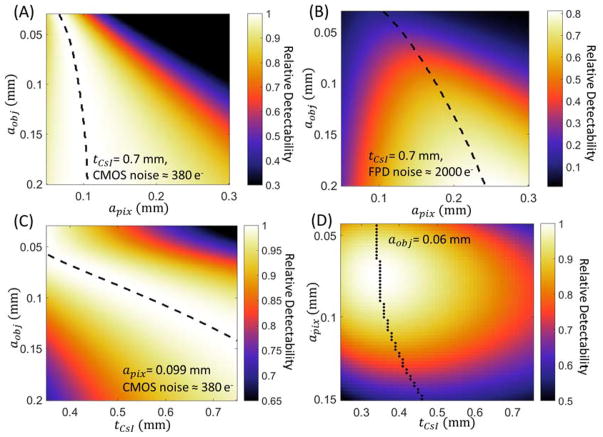
Task-based evaluation of CMOS detector performance in extremity CBCT. (A) Relative detectability for a range of feature sizes (vertical axis) as a function of pixel size. Scintillator thickness is assumed constant and equal to 0.7 mm. Detectability is normalized to the maximum value for each aobj. Dashed lines indicate maximum d′ for each feature size. (B) Ratio of d′2 achieved with the same scintillator as (A), but at increased electronic noise consistent with an a-Si:H FPD, to maximum d′2 attained for each aobj by the low-noise CMOS detector of (A). (C) Relative detectability of the CMOS detector as a function of scintillator thickness and imaging task, normalized by maximum detectability achieved for each aobj across the range of tCsI. Pixel size is 0.099 mm. (D) Joint optimization of pixel size and scintillator thickness for a “trabecular” imaging task with feature size of 0.06 mm. The graph shows detectability of a CMOS detector (normalized by the maximum).

