Figure 7.
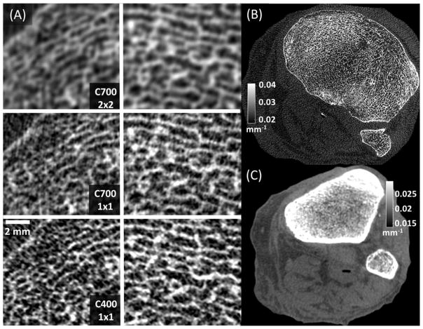
(A) Magnified views of two trabecular regions in the subchondral bone of a cadaver knee imaged using CMOS detectors with different pixel sizes and scintillator thicknesses. High resolution bone reconstruction was used. (A, top) Reconstructions of 2×2 binned C700 projections, mimicking the pixel size of current a-Si:H FPDs. (A, middle) Reconstructions of C700 projections in 1×1 binning, showing the benefits of reduced pixel size provided by CMOS. (A, bottom) Images acquired with C400 in 1×1 binning, illustrating the visualization of trabecular detail using a thin scintillator. (B) A complete axial slice of C400 reconstruction obtained using high resolution protocol (C) A C400 reconstruction obtained using a soft-tissue protocol with 4×4 pixel binning.

