Figure 4.
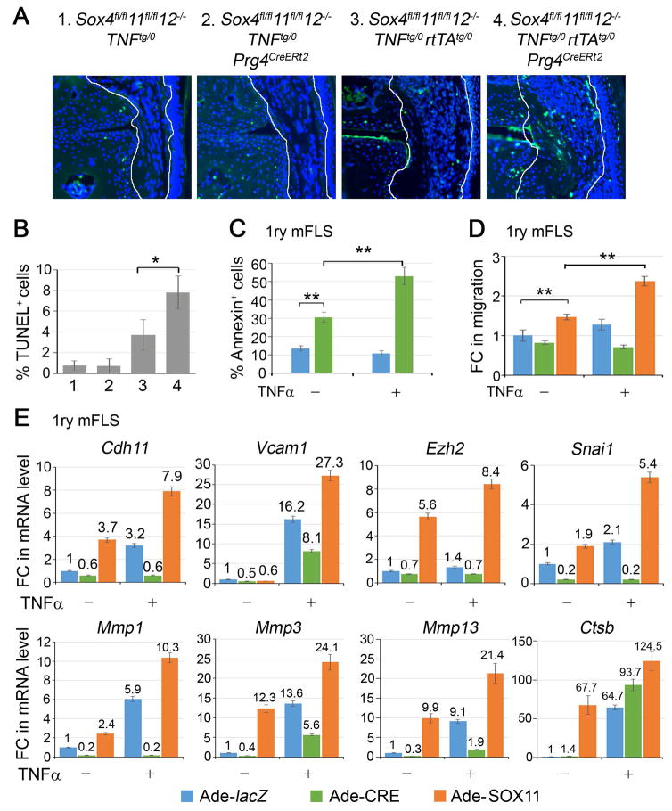
SOXC genes promote FLS transformation. A, TUNEL staining (green) and DAPI counterstaining (blue) of cell nuclei. B, The graph shows the percentage of TUNEL-positive synovial cells from 3 mice for genotype. C, Flow cytometric quantification of Annexin V-positive cells in Sox4fl/fl11fl/fl12−/− primary FLS infected with lacZ- or CRE-expressing adenovirus for 16 h and then treated with 5 ng/ml TNFα for 8 h. D, Assay of wound repair by primary FLS. Confluent cultures of Sox4fl/fl11fl/fl12−/− FLS were infected with lacZ-, CRE- or SOX11-expressing adenovirus for 16 h, wounded with a pipette tip, and then treated or not with 5 ng/ml TNFα for 4 h. E, Fold changes in the relative levels of RNAs for the indicated genes in Sox4fl/fl11fl/fl12−/− primary FLS treated adenoviruses for 16 h and then with 5 ng/ml TNFα for 8 h, as indicated. Gapdh mRNA levels were used for normalization.*, p-value <0.05; **, p-value <0.01 in two-tailed Student’s t-test applied to triplicate cultures per condition.

