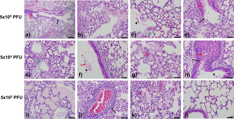
Figure 4. Mouse-adapted M35c4 elicits pathology associated with ARDS.
Representative images were acquired from mouse lungs infected with maM35c4 at doses of 5×10⁵ (a–d), 5×10⁴ (e–h), or 5×10³ (i–l) PFU at day 7 p.i. Images reflect the severity of disease observed at the different doses including denuding of large airways (a, d, h), debris in the airways (a, d, f, h), hyaline membranes (a–e), red blood cells in the large airways and alveoli (c, f, h), edema (c, d, g, I, k), and perivascular cuffing (j). A normal region of a mouse lung at the 5×10³ PFU dose is shown (l). Image (a) was captured at 10× magnification and all other images were acquired at 40× magnification. Images are representative of at least 3 samples. Scale bars in lower right corners of each panel are 1mm.

