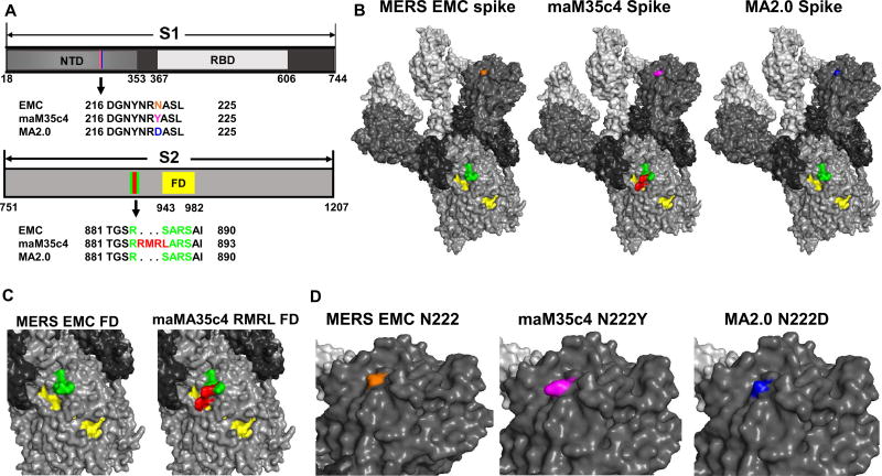
Figure 5. Structural modeling of the maM35c4 spike mutations against EMC 2012 and MA2.0.
a. Alignment of the amino acid sequences identified in the S1 and S2 domains from EMC 2012, maM35c4, and MA2.0. The colors in S1 correspond to amino acid sequence changes in the N-terminal domains (NTD) of maM35c4 (magenta, N222Y), and MA2.0 (blue, N222D) compared to EMC 2012 (orange). In the S2 domain the RMR insertion and S885L mutations in maM35c4 (red) are compared to the wild-type sequence in EMC 2012 and MA2.0 (green). The S2’ fusion domain region is highlighted in yellow. Varying shades of light gray to black in the S1 and S2 diagrams correspond with the same coloring scheme depicted for the spike trimers in (b). b. Structural modeling of MERS-CoV monomers was conducted using the PHYRE2 online modeling software; trimers were constructed using the PyMOL Molecular Graphics System. All the respective mutations identified within the spikes of maM35c4 and MA2.0 were incorporated into the modeled structure. The additional mutations in MA2.0 are not visible on the surface of the structure as shown. c and d. Expanded views of the structural regions observed near the S2’ and fusion domains of EMC 2012 and maM35c4 (c) and the region of the NTD encompassing amino acid N222 from EMC 2012, maM35c4, and MA2.0 (d).

