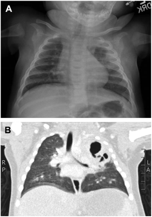
Fig. 1.

Radiographic imaging in cavitating pneumonia. (A) Chest radiograph demonstrating a complex air space opacity in the left upper lobe with central lucency consistent with cavitating pneumonia. (B) CT of the same lesion demonstrates a large cavity with central necrosis and multiple air fluid levels occupying most of the left upper lobe.
