Figure 2.
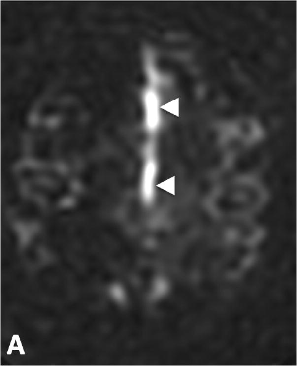
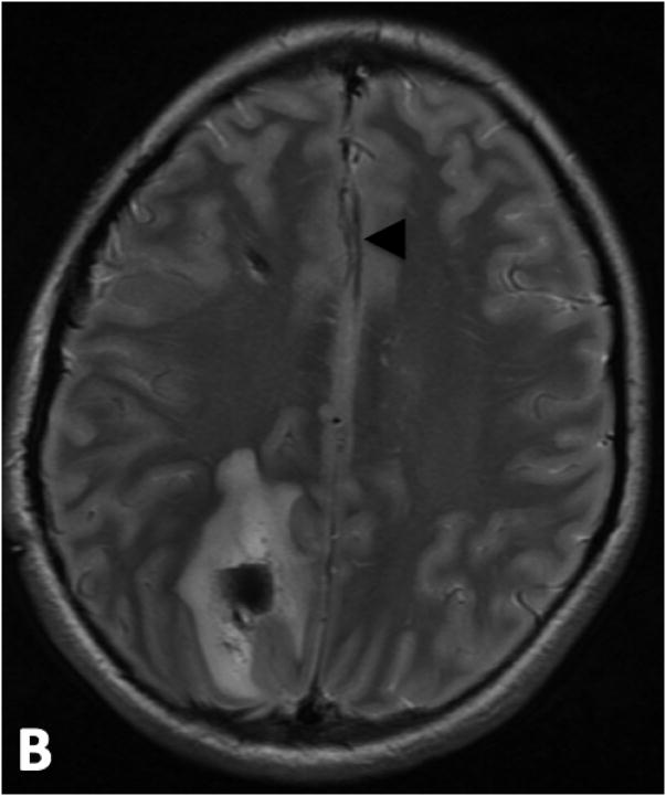
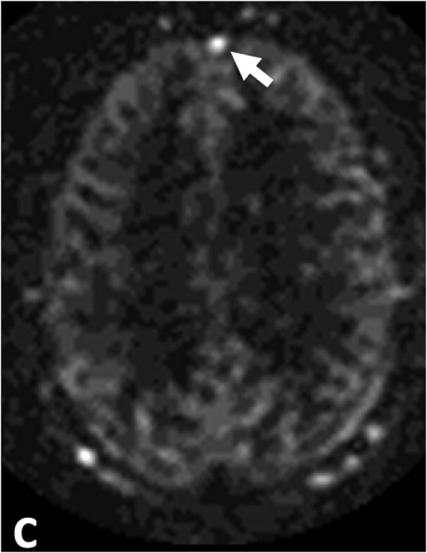
False positive venous ASL signal in two patients. A. ASL signal in the A3 branches of the anterior cerebral arteries (white arrow heads) was mistaken for venous ASL signal in a 15 year-old male patient who presented with a right parietal parenchymal hematoma. B. T2-weighted images show localization of this signal to the anterior cerebral arteries (black arrow heads). C. ASL signal in the anterior aspect of the superior sagittal sinus in a 70 year-old male with subarachnoid hemorrhage (white arrow). This patient had no evidence of a dAVF or shunting on DSA.


