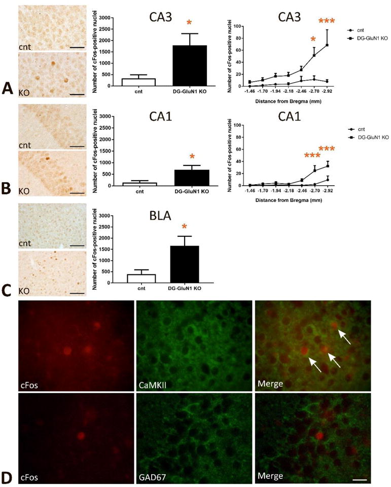
Figure 3.

(A) Increased number of cFos-positive nuclei in hippocampal CA3. Left panel, representative images in cont and DG-GluN1 KO brains. Middle panel, the total number of cFos-positive nuclei was significantly increased in CA3 (t(8)=2.665, * p=0.02). Right panel, the number of cFos-positive nuclei over the rostral (dorsal)-caudal (ventral) axis of CA3 of the hippocampus (−1.46 to −2.92 mm from Bregma). 2-Way ANOVA analyses show significant genotype, hippocampal rostral-caudal axis and genotype X hippocampal rostral-caudal axis interaction in CA3 (Genotype effect: F(6, 56)=4.635, *** p=0.0007; hippocampal rostral-caudal axis effect: F(1, 56)=24.53, **** p<0.0001; and interaction effect: F(6, 56)=2.634, * p=0.0254). Post-hoc comparison further demonstrates significant increased cFos-positive nuclei in the caudal (ventral) CA3 (* p=0.01 at −2.70mm from Bregma, **** p<0.0001 at −2.92mm from Bregma).
(B) Increased cFos-positive nuclei in hippocampal CA1. Left panel, representative images. Middle panel, increased total number of cFos-positive nuclei in CA1 (t(8)=2.614, * p=0.03). Right panel, the number of cFos-positive nuclei over the rostral-caudal axis of CA1 of the hippocampus (−1.46 to −2.92 mm from Bregma). 2-Way ANOVA analyses show significant genotype, hippocampal rostral-caudal axis and genotype X hippocampal rostral-caudal axis interaction in CA1 (Genotype effect: F(6, 56)=8.286, **** p<0.0001; hippocampal rostral-caudal axis effect: F(1, 56)=18.34, **** p<0.0001; and interaction effect: F(6, 56)=3.374, ** p=0.0066). Post-hoc comparison further demonstrates significant increased cFos-positive nuclei in the caudal CA1 (*** p=0.0007 at −2.70mm from Bregma, *** p=0.0005 at −2.92mm from Bregma).
(C) Increased number of cFos-positive nuclei in basolateral amygdala (BLA, −0.70 to −2.30 mm from Bregma, t(8)=2.709, * p=0.02). Left panel, representative images.
(D) The majority of cFos-positive nuclei in hippocampal pyramidal layer in CA3 subfield were located within CaMKII-positive excitatory neurons, but not within GAD-67-positive inhibitory neurons.
