Figure 5.

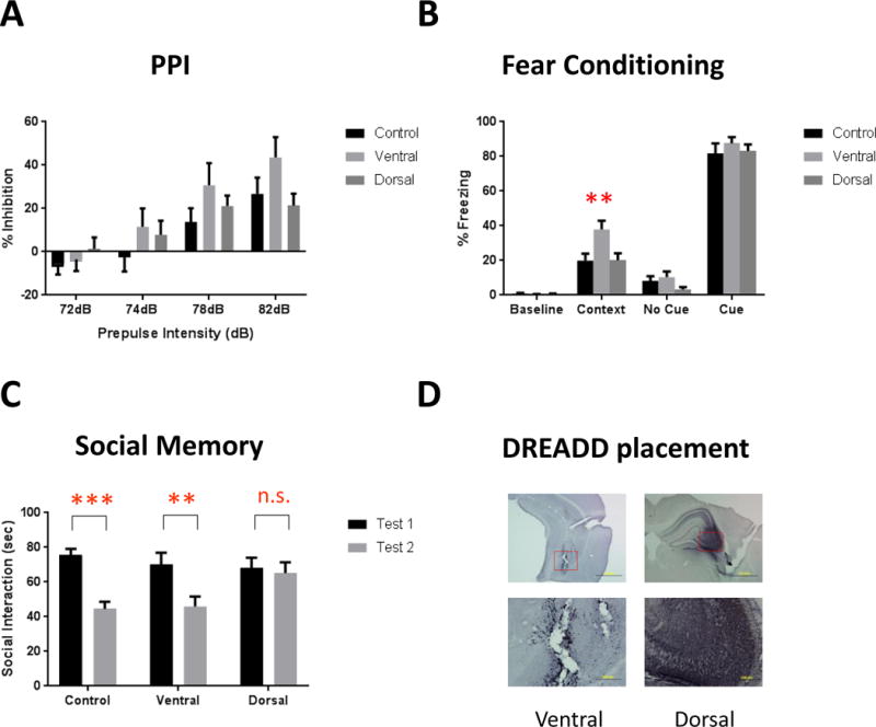
Behavioral analysis after DREADD-induced excitation of CA3. (A) Prepulse inhibition. 2-way ANOVA revealed a main effect of prepulse intensity (F(3,81)=42.08, p<0.0001) and a prepulse × group interaction (F(6,81)=2.224, p=0.049). However, post-hoc analysis shows no difference between any group at any specific prepulse intensity.
(B) Fear conditioning. There is a significant main effect of region (F(2,103)=4.887, p=0.0094), and post-hoc analysis revealed a significant increase in contextual fear conditioning after activation of the ventral CA3 (p=0.0016). Activation of this region did not affect baseline freezing or cued fear, and activation of the dorsal CA3 did not alter any fear-related behavioral measure.
(C) Social memory. Normal social recognition is demonstrated by a significant decrease in interaction time between the first and second tests and was present in control animals (t=4.237, df1,54, p<0.001). Ventral CA3-activated DREADD mice significantly decreased interaction time (t=2.994, df1,54, p<0.01), whereas activation of the dorsal CA3 with the excitatory DREADD impaired this recognition, resulting in no decrease in interaction time between the two tests (t=0.3649, df1,54, p=0.717).
(D) Verification of DREADD placement based on mCherry staining. DAB signaling was enhanced by the addition of nickel sulfate to give the mCherry staining a black color, distinguishing the specific mCherry signal from background gliosis present as a result of the AAV infusion. Images represent the ventral (left) and dorsal (right) CA3 at 4× (top) and 20× (bottom). **, *** represents p<0.01, 0.001, respectively.
