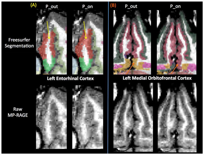
Figure 8.
Segmentation differences between human MP-RAGE P_on (pump in the room and on) and P_out (no pump or cable in room) for two regions with percentage differences in volume estimates greater than 5%, the left entorhinal cortex (a) and the left medial orbitofrontal cortex (b). Freesurfer segmentations shown in the top panes and raw MP-RAGEs shown in the bottom panes. (a) Left entorhinal cortex (red): Yellow arrows indicate an area of white matter incorrectly segmented as left entorhinal cortex only in P_on. (b) Left medial orbitofrontal cortex (pink): Orange arrows indicate an area in P_on that is incorrectly segmented as left medial orbitofrontal cortex instead of left insula (yellow).

