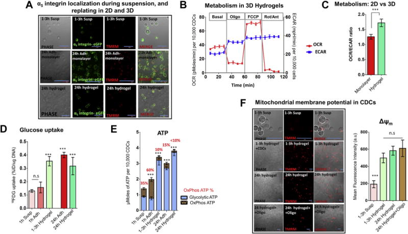
FIGURE 6. Energetics and Glycolytic Reserve Are Restored Following CDC Encapsulation in HA:Bl:Ser Hydrogels.

(A) Integrin localization. Cell plating as 2D monolayers and encapsulation in HA:Bl:Ser hydrogels (but not cell suspension) leads to α5-integrin localization in the cell membrane within 1 h, indicating activation of cell adhesion. Calibration bar in microscopy figures represents 50 μm. (B and C) Respirometry. Cell encapsulation in hydrogels leads to a rapid increase in OCR and ECAR. The OCR/ECAR ratio of cells encapsulated in hydrogels is higher than cells cultured as adherent monolayers. Statistical comparison was performed using 1-way ANOVA followed by Tukey’s post-hoc test. (D and E) Glycolytic reserve. Glucose (18FDG) uptake and glycolytic ATP generation are restored within 1 to 3 h of encapsulation in hydrogels. Statistical comparison was made using the Student t test with the 1-h suspension condition. (F) Mitochondrial membrane potential. ΔΨm, an indicator of cell viability, is maintained after inhibition of OxPhos by oligo (24 h), indicating presence of glycolytic reserve in cells encapsulated in hydrogels. Statistical comparison was made using the Student t test. against the 1- to 3-h suspension condition. Calibration bar represents 50 μm; n = 3. Hydrogels refers to HA:Bl:Ser hydrogels. Data is presented as mean ± SD. ***p < 0.001. Abbreviations as in Figure 1.
