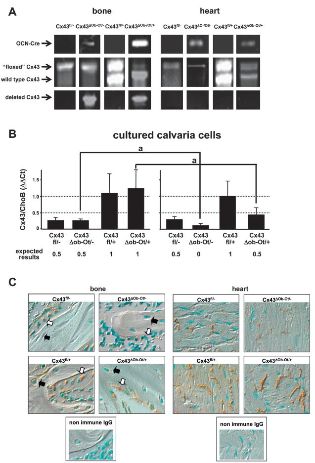
FIG. 1.
Genotypic and phenotypic analysis of mice lacking Cx43 in osteocytes and osteoblasts and their littermates. (A) Genomic DNA was purified from mouse bone (tibia) and heart and PCR for OCNCre, “floxed,” and wildtype Cx43 allele and deleted Cx43 were performed. (B) Calvaria cells were isolated from mice carrying the four different genotypes and treated with ascorbic acid for 0 or 21 days. The levels of Cx43 mRNA were determined by real-time PCR and corrected by ChoB. The expected levels of expression of Cx43 are indicated. *p < 0.001 vs. day 0, n = 3–9 mice. (C) Representative microphotographs of paraffin-embedded bone (tibia) and heart sections immunostained for Cx43 (brown) and counterstained with methyl green to show the cell nuclei (blue nuclei). An example of an osteocyte in each bone section is pointed out by black arrows and an example of an osteoblast by white arrows.

