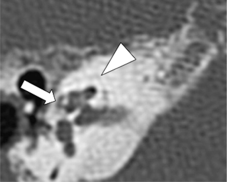
Fig 3.

Axial CT shows presence of a cavitary plaque involving the pericochlear region (arrow), note the density of the cavitary plaque similar to the components in the IAC. Additionally, there are noncavitary otospongiotic plaques surrounding the otic capsule (arrowhead).
