
Figure 1.
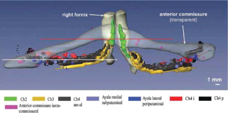
Anatomy of the basal forebrain complex. 3D-reconstruction of the basal forebrain complex (BFC–view from anterior) from the brain of a 29-year-old man who had died of pulmonary arrest [Grinberg and Heinsen, 2007]. The BFC is located within the substantia innominata that is delimited by the caudal rim of the ventral striatum, the ventral pallidum, the ventral parts of the internal capsule and the regions medial to the outlines of the anterior commissure. The BCF can be subdivided into four cell groups arranged in an arch-like path mainly beneath the anterior commissure: Ch1 or medial septal nucleus; Ch2 and Ch3 or the nucleus of vertical and horizontal limb of the diagonal band of Broca, respectively; and Ch4 also called as the nucleus basalis of Meynert [Mesulam et al., 1983]. The nucleus subputaminalis, also called Ayala’s nucleus, has only been described in the human brain so far [Heinsen et al., 2006; Simic et al., 1999]. The volume of the BFC in the human brain varies from 58 to 154 mm3 [Grinberg and Heinsen, 2007; Halliday et al., 1993].