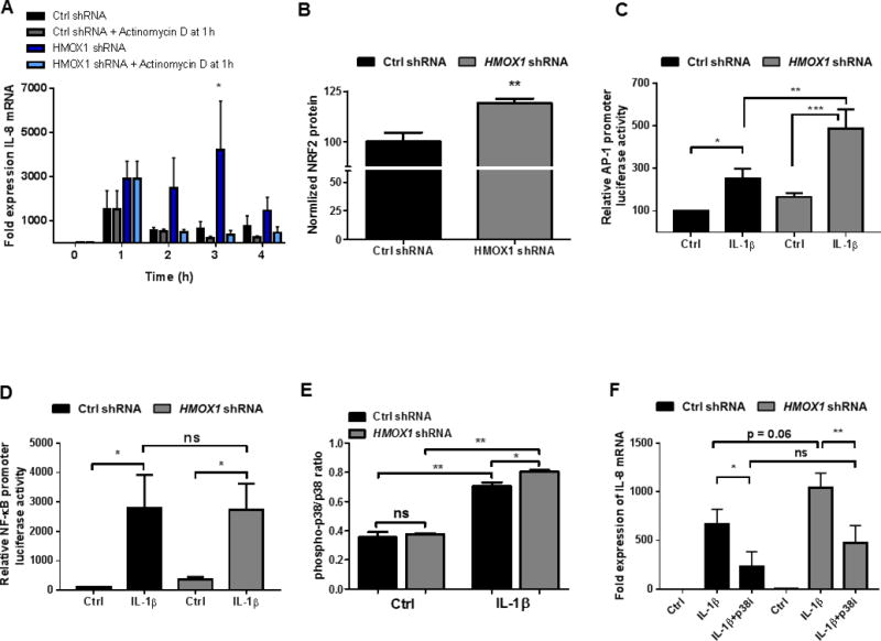
Figure 5. Mechanisms of transcriptional regulation by HO-1.

(A) Expression of IL-8 in confluent NTC shRNA (Ctrl) and HMOX1 shRNA transduced Caco-2 IECs treated with IL-1β (1 ng/mL) +/− transcriptional blockade with Actinomycin D after 1h. (B) Expression of NRF2 protein at baseline normalized to cell density via Crystal Violet staining and presented as % of control. Activation of AP-1 (C) and NF-κB (D) transcription factors by IL-1β (1 ng/mL) was measured using luciferase reporter plasmid transfection in NTC shRNA and HMOX1 shRNA transduced Caco-2 IECs. (E) Activation (phosphorylation) of p38 MAPK was assessed at baseline and in the setting of IL-1β treatment (1 ng/mL × 30 minutes) by cell-based ELISA. (F) The influence of inhibition of p38 MAPK on the increased expression of IL-8 in HMOX1 shRNA Caco-2 cells was assessed by pretreatment with a p38 MAPK inhibitor (SB202190 at 40 μM) 1h prior to IL-1β (1 ng/mL) exposure for 2h. Data represent combined results from at least 3 independent experiments. *, p <0.05; **, p <0.01.
