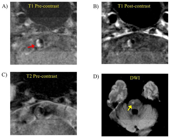
Figure 1.
Intraplaque hemorrhage presented in a low grade stenotic basilar artery plaque (43% degree of stenosis) in an acute symptomatic patient (age 65, female). A) T1 weighted black blood MRI showed high signal (fresh IPH, red arrow) in the plaque. B) Post contrast T1 weighted images showed slight enhancement of the plaque. C) T2 weighted images showed iso-intense signal of the plaque. D) DWI showed infarct in the brain stem (yellow arrow).

