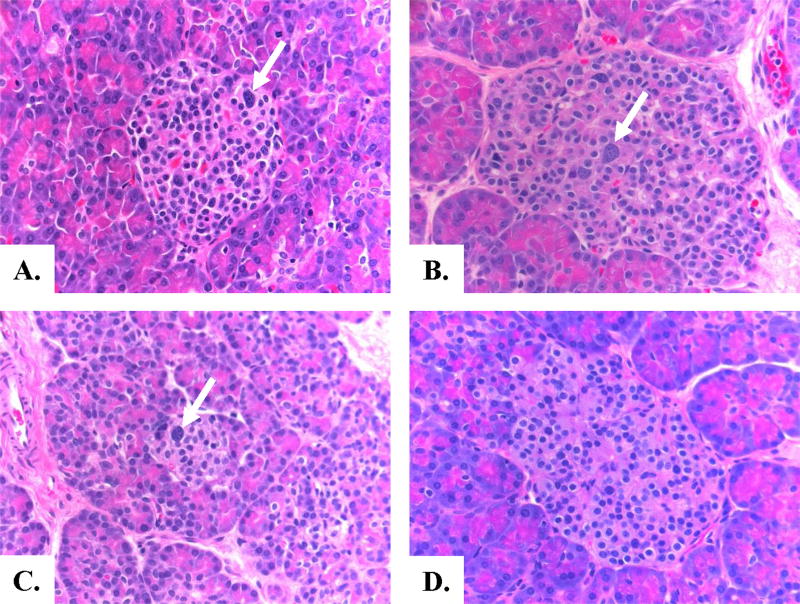
Figure 1. Pancreatic Histology of Congenital Hyperinsulinism in Turner Syndrome.
Panels A and B: Appearance of pancreatic islets in Case 3 and Case 7 with Turner Syndrome and Hyperinsulinism. Panel C: Pancreas of infant with diffuse KATP hyperinsulinism. Panel D: Normal pancreas from a control infant. Histopathology of the two Turner Syndrome cases and the KATP hyperinsulinism case show similar changes of scattered islet cell nucleomegaly (arrows)and normal lobular parenchymal architecture typical of diffuse hyperinsulinism. (H and E staining, 40× magnification)

