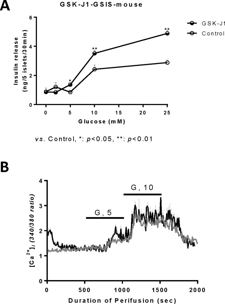
Figure 4. Effects of a KDM6A inhibitor (GSK-J1) on responses of normal isolated mouse islets.
Isolated mouse islets were first cultured with KDM6A inhibitor (GSK-J1), 0.5 µM, for 3 days, removed from GSK-J1 during a 30-minute glucose-free pre-incubation, and then exposed to different concentrations of glucose for another 30 minutes in the absence of inhibitor. Panel A: Islets incubated with GSK-J1 (filled circles) exhibited a leftward shift in glucose-stimulated insulin secretion with increased release of insulin at 10 mM and 25 mM glucose compared to control islets (open circles). Panel B: Basal cytosolic calcium was initially elevated in islets following exposure to GSK-J1 inhibitor (black line) during 3 days of culture, but showed similar cytosolic calcium responses compared to control islets (grey line) during perifusion with 5mM or 10mM glucose in the absence of GSK-J1.

