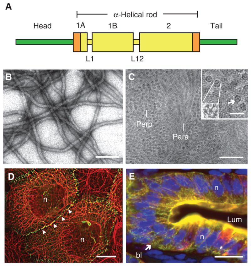
Figure 1.

Introduction to keratin intermediate filaments (IFs). (A) Schematic representation of the tripartite domain structure shared by all keratin and other IF proteins. A central α-helical “rod” domain acts as the major determinant of self-assembly and is flanked by nonhelical “head” and “tail” domains at the amino and carboxyl termini, respectively. Within this 310-amino-acid-long rod domain, the heptad-repeat-containing segments (e.g., coils 1A, 1B, and 2) are interrupted by linker sequences at two conserved locations (e.g., L1 and L12). Rod domain boundaries consist of highly conserved 15–20-amino-acid regions (shown in orange) that are crucial for polymerization and frequently mutated in human disease (see www.interfil.org). (B) Visualization of filaments, reconstituted in vitro from purified human K5 and K14 proteins, by negative staining and electron microscopy. Scale bar, 100 nm. (C) Visualization of native keratin IFs in the stratum corneum layer of human epidermis using cryo-transmission electron microscopy on a fully hydrated, vitreous tissue specimen (Norlén and Al-Amoudi 2004). Bundles of tightly packed keratin IFs run parallel (para) or perpendicular (perp) to the sectioning plane. Scale bar, 50 nm. (Inset) Detailed views of filaments in cross section, shown at two magnifications. As many as seven subfibrils, including a centrally located one, can be seen. Scale bar, 20 nm. (D) Skin epidermal keratinocytes in culture. Dual-immunofluorescence labeling for keratin (red signal) and desmoplakin (green signal), a desmosome component. Keratin IFs are organized in a network that spans the whole cytoplasm and are attached to desmosomes, which are points of adhesion at cell–cell contacts. n, Nucleus. Scale bar, 20 μm. (E) Gut epithelial wall in cross section, emphasizing the epithelium. This fresh-frozen specimen was triple-labeled for K8/K18 (red signal), K19 (green signal), and nuclei (blue signal). Note the concentration of staining at the apical pole of enterocytes. The star denotes a goblet cell, which also features a prominent K8/K18 network in the cytoplasm. bl, Basal lamina; lum, lumen; n, nucleus. Scale bar, 20 μm. (Reprinted, with permission, from Kim and Coulombe 2007; C, originally adapted from Norlén and Al-Amoudi 2004, with permission from Elsevier.)
