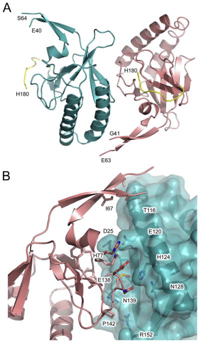
Fig. 2.
hHRF-1. (A) Overall structure of hHRF-1 solved at 1.75 Å resolution. The two molecules of the asymmetric unit are coloured blue and salmon. The C-terminal tag is coloured yellow and the positions of C-terminal residues, and residues adjacent to the disordered loop, are indicated. (B) The two monomers of the asymmetric unit (blue and salmon) bury a surface area of ~530 Å2 at their interface. (For interpretation of the references to colour in this figure legend, the reader is referred to the web version of this article.)

