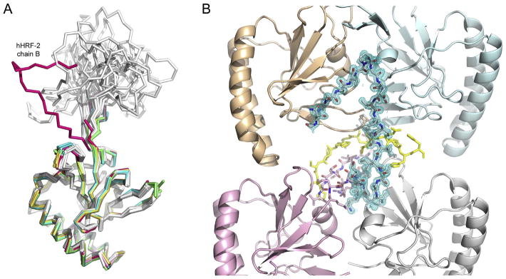
Fig. 4.
The hHRF mobile loop. (A) In chain B of the hHRF-2 structure, the mobile loop conformation differs from that in the hHRF solution structure, and partially ordered loops in other hHRF crystal structures. Structures are coloured as follows: hHRF NMR structure (PDB 2HR9, 20 conformers), grey (Feng et al., 2007); hHRF crystal structure (PDB 1YZ1, four chains), green (Susini et al., 2008); hHRF crystal structure (PDB 3EBM, four chains), yellow (Dong et al., 2009); hHRF-1 structure chain A, pale blue; hHRF-1 structure chain B. teal; hHRF-2 structure chain A, pale pink; hHRF-2 structure chain B, dark pink. For clarity, C-terminal tags are not shown. (B) The mobile loop from chain B of the hHRF-2 structure packs against a symmetry-related dimer. Chains A and B of the hHRF-2 structure are coloured pink and grey, respectively, and loop residues are shown as sticks. Electron density is shown for the loop from chain B (2FoFc map contoured at 1σ). Symmetry-related molecules are coloured in pale orange and blue, and the C-terminal tag is coloured yellow. (For interpretation of the references to colour in this figure legend, the reader is referred to the web version of this article.)

