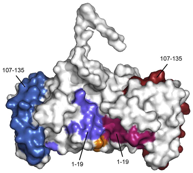
Fig. 6.

The hHRF-2 disulphide-linked dimer is consistent with a model of mast cell activation. The two monomers of the hHRF-2 dimer are coloured white and Cys172 is coloured orange. For the first monomer, the two IgE binding sites, mapped to residues Met1-Lys19, and Arg (hHRF)/Lys (mHRF) 107-Ile135, are coloured light blue and dark blue respectively. For the second monomer, residues 1–19 and 107–135, are coloured light pink and dark pink respectively. This dimeric structure offers a model not only for human, but also murine, HRF in mast cell activation. (For interpretation of the references to colour in this figure legend, the reader is referred to the web version of this article.)
