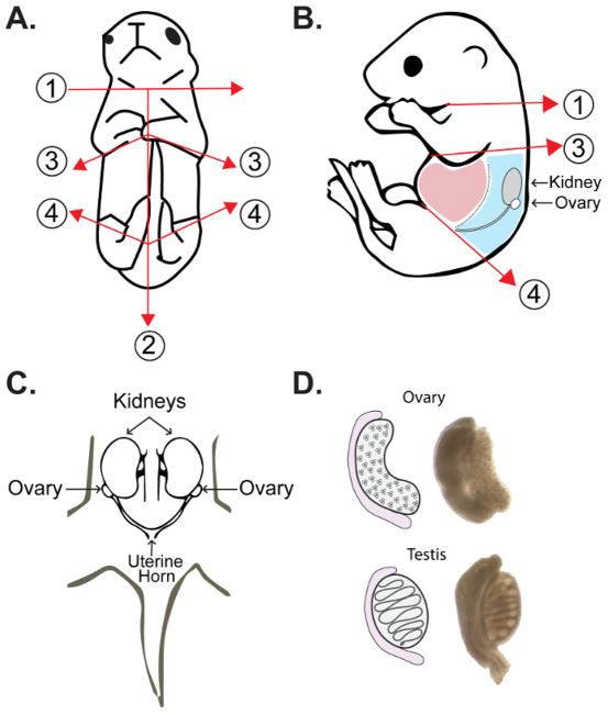
Figure 2. Ovary extraction from embryos and neonatal female pups.
(A, B) The first cut (1) is made above the forelimbs to decapitate the embryo at the head/neck junction immediately upon retrieval from the maternal uterine horn. The second cut (2) is made along the ventral midline of the posterior half of the embryo, followed by incisions along the anterior half below the forelimbs (3). A final cut is made for removal the hind limbs and tail (4). (A) Frontal view schematic of dissection cuts for isolation of ovaries from female pups. (B) Side view schematic of dissection cuts for isolation of ovaries with relative positions of internal organs. Regions shown in light red include the liver and intestines, which are removed during dissection. The dorsal wall and associated organs are shown in light blue. This region includes the ovaries, which are attached to the inferior poles of the kidneys at the top of the uterine horn. (C) Frontal schematic view of dorsal wall of embryo following removal of the liver and intestines. (D) Schematic representation of morphological differences between male and female gonads at approximately 15–18 dpc. Please click here to view a larger version of this figure.

