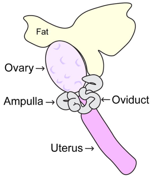
Figure 4. Schematic diagram of adult ovary.
This diagram depicts the anatomy of the adult mouse ovary, oviduct, ampulla and uterus. Mouse ovaries should be removed by careful incision of ligaments connecting the ovaries to the inferior poles of the kidneys, and the posterior wall of the abdomen. Please click here to view a larger version of this figure.

