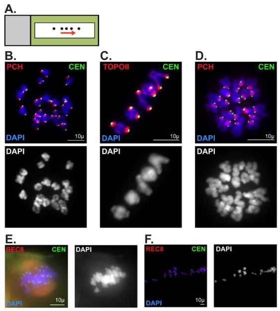
Figure 6. Representative metaphase I and II oocyte chromatin spread preparations.
(A) Slide schematic for metaphase I and II oocyte chromatin spread preparations. Oocytes (circles) released via mouth-operated glass pipette or capillary in a straight line (following the arrow). Black rectangle outline represents liquid blocker pen outline. (B, C) Optimal metaphase I chromatin spread preparation. Metaphase I chromosomes were stained with DAPI (blue, DNA), and immunolabeled for CEN (green, kinetochore/centromere marker). Scale bars = 10 μm. (B) Antibodies against Histone H4 (di methyl K20, tri methyl K20) were used to label pericentromeric heterochromatin (PCH). (C) Antibodies against Topoisomerase IIα (TOPOII) were used to label the PCH and chromosome axes. (D) Optimal metaphase II chromatin spread preparation. Metaphase II chromosomes were stained with DAPI (blue, DNA), and immunolabeled for CEN (green), and Histone H4 (di-methyl K20, tri-methyl K20) to label the PCH. Scale bar: 10μm. (E, F) Poor metaphase I chromatin spread preparations. Chromosomes were stained with DAPI (blue, DNA), and immunolabeled for CEN (green) and the meiotic cohesin component, REC8. Scale bars = 10 μm (E) An oocyte that did not burst upon release onto PFA-coated slide. (F) Chromosomes that were spread too far apart. Please click here to view a larger version of this figure.

