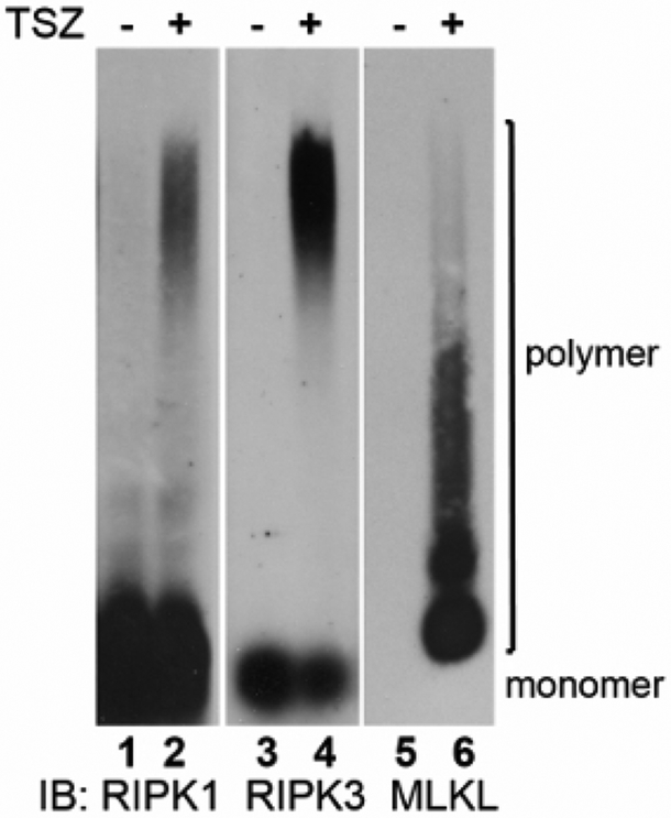
Figure 1. Examination of amyloid-like fibers during necroptosis.
Whole cell lysates were harvested from cells undergoing TNF-induced necroptosis and subjected to SDD-AGE. Western blots were performed for RIPK1, RIPK3, and MLKL. The MLKL-containing fibers exhibited a distinct migration pattern from the RIPK1/RIPK3-containing fibers. The MLKL monomer is barely visible under this condition.

