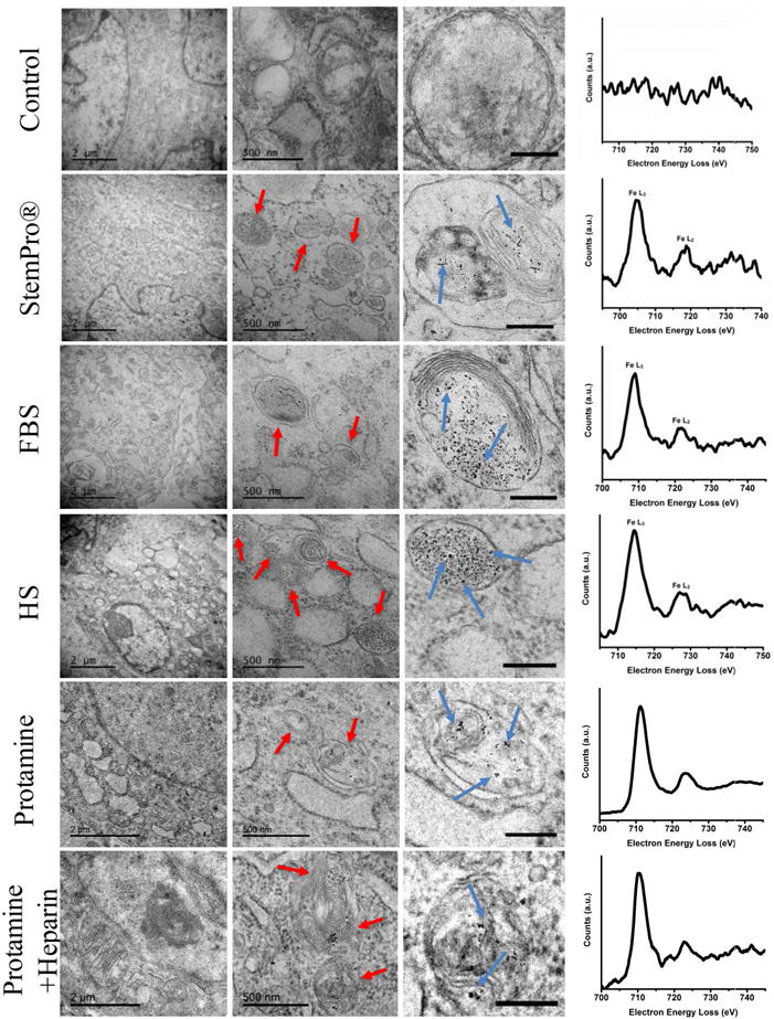
Figure 4.

Ferumoxytol compartmentalization in hMSC after exposure to different labeling media: Electron microscopy of similarly treated cells shows nanoparticle-containing lysosomes/endosomes (red arrows) and the iron nanoparticles (blue arrows) in all samples that have been exposed to ferumoxytol (scale bar on left column, middle column, and right column are 2μm, 500nm, and 200nm respectively). Electron energy loss spectra (right) confirm the presence of iron in lysosomes/endosomes of labeled cells and the absence of iron in the unlabeled control.
