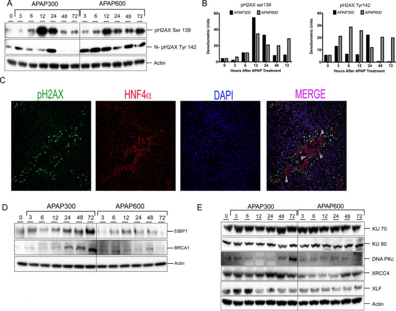
Figure 2.

Prolonged DNA DSB and reduced repair protein expression after higher dose of APAP. (A) Western blot analysis of phos-H2AX Ser139 using total liver extract and phos-H2AX Tyr142 using nuclear extract. (B) Bar graphs showing densitometric analysis of pH2AX ser139 and Tyr142 Western blots (C) Representative immunofluorescence staining for pH2AX Ser139 (green), HNF4α (red) and DAPI (blue) for cell nuclei. Arrowheads are pointing to necrotic cells. (D) Western blot analysis of DNA repair mediator proteins 53BP1, BRCA1in total liver extract (E) Western blot analysis of DNA repair effector proteins KU70, KU80, DNAPkc, XRCC4, XLF, Lig4 using total liver extract.
