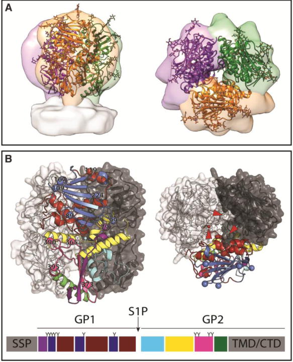
Figure 1. The unusual arenavirus GP trimer.

(A) The crystal structure of the LASV GP trimer (cartoon, PDB: 5VK2) docked into the tomographic reconstruction of the LASV GPC spike from fixed virions (surface, EMD: 3290). Glycans visible in the crystal structure are shown as ball and stick. (B) GP monomer A is shown in cartoon representation. LASV GP1 can be divided into three sub-domains: (1) the N-terminal β-strands (purple, interacting with GP2); (2) the upper, β-sheet core (blue); and (3) the lower helix-loop surface (dark red). LASV GP2 can be divided into four sub-domains: (1) the fusion region (cyan), which is composed of the fusion peptide and fusion loop; (2) HR1a-d (yellow); (3) the T-loop (magenta); and (4) HR2 (green). N-linked glycosylation sites are shown as spheres and numbered on their respective Asn residue. GP monomers B and C are shown as surfaces. The C terminus of each GP1 subunit is indicated with an arrow and the N termini of the GP2 subunits, which are positioned at the three-fold axis and 30Å below the apex of the trimer, are indicated with a star. A linear representation of the GPC protomer is shown a bottom with each subdomain of GPC colored according to the cartoon representation, with each N-linked glycosylation site indicated with a “Y”.
