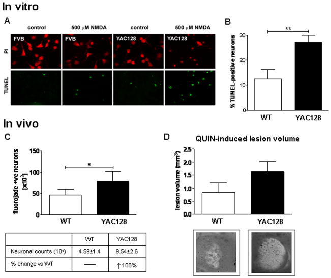
Fig. 1. Increased sensitivity to excitotoxic stress in YAC128 mice before signs of illness.
(A and B) Primary neuronal cultures established from embryonic YAC128 (HD53) and WT striata were assessed for apoptotic cell death 24 hrs post NMDA. MSNs from YAC128 (HD53) striata demonstrate an increase in vulnerability to NMDAR-mediated excitototxicity versus WT with increased cell death observed post NMDA (p<0.01). C) Intrastriatal injection of QUIN in 1.5 month old mice demonstrates an increase in apoptotic Fluoro-Jade positive cells in YAC128 (HD53) striatum compared to WT (p<0.05). D) Quantification of lesion volume demonstrates a trend towards enhanced lesion volume in YAC128 (HD53) compared to WT striata (p<0.07). Lesion volume and mean number of Fluoro-Jade positive cell is ± SEM. Mean percent apoptotic cell death is given ± SD. *p<0.05; **p<0.01.

