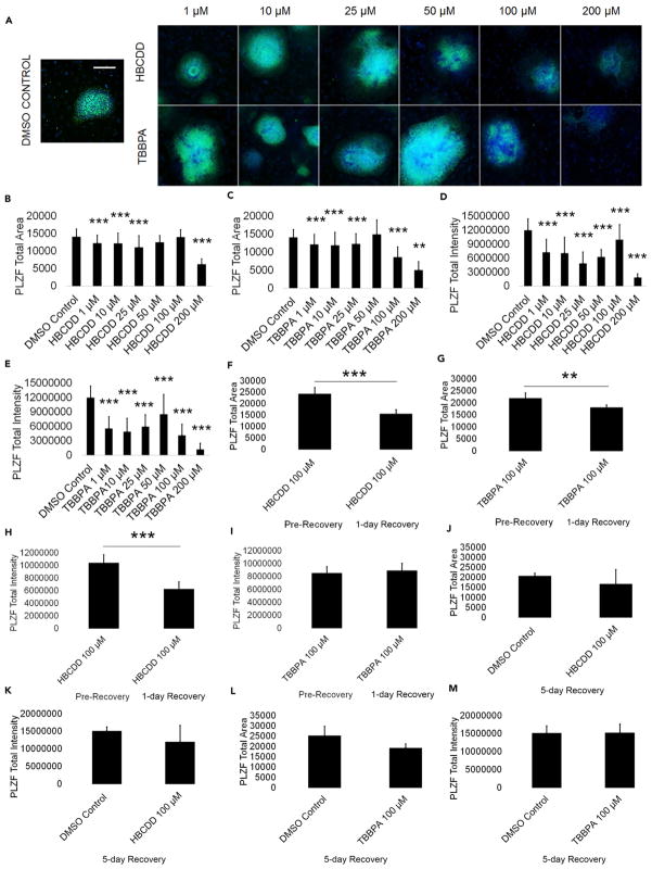
Figure 2. HBCDD and TBBPA Reduce PLZF+ Spermatogonia in In Vitro Spermatogenic Cultures.
(A) Representative 5X images of PLZF+ (green) and DAPI (blue)-stained colonies treated with HBCDD and TBBPA. A control image is included; scale bar, 5,000 μm. All images are taken under the same imaging conditions and parameters.
(B and C) Graphical representation showing that HBCDD (B) and TBBPA (C) reduce the average total PLZF+ area in spermatogonia derived under in vitro spermatogenic conditions.
(D and E) Graphical representation showing that HBCDD (D) and TBBPA (E) reduce the average total PLZF+ intensity in spermatogonia.
(F and G) Graphical representation showing that HBCDD (F) and TBBPA (G) exposure continues to reduce the average total PLZF+ area in spermatogonia, even after a 24-hr chemical-free recovery period.
(H and I) Graphical representation showing that HBCDD (H) and TBBPA (I) exposure continues to reduce the average total PLZF+ intensity in spermatogonia, even after a 24-hr chemical-free recovery period.
(J–M) Graphical representation showing that spermatogonia are capable of recovery following a 5-day recovery period after 100 μM TBBPA and HBCDD exposure. Five replications were performed for each condition for (B)–(E). Three replications were performed for each condition for (F)–(M). Significant changes in PLZF+ area and intensity were determined using a 1-way analysis of variance (ANOVA) and validated via a Student’s t test, where ** is p < 0.01, and *** is p < 0.001. Data are represented as mean ± SEM.
See also Figure S2.

